Doublechambered right ventricle a commonly overlooked diagnosis HKMJ
Double-chambered right ventricle is a congenital anomaly in which the right ventricle is divided into 2 portions by anomalous muscle bundles. These cases often present in children, but rarely in adults. We discuss 2 cases of double-chambered right ventricle, in patients aged 42 and 35 years.

[PDF] Doublechambered right ventricle or subinfundibular stenosis
Double-chambered right ventricle is a rare congenital heart disorder involving 2 different RV pressure compartments that is often associated with malalignment VSD. Usually, the obstruction is caused by an anomalous muscle bundle crossing the RV from the interventricular septum to the RV free wall.

Double Outlet Right Ventricle Repair, Surgery & Survival Rate
Double-chambered right ventricle is a rare congenital heart disorder involving 2 different RV pressure compartments that is often associated with malalignment VSD. Usually, the obstruction is caused by an anomalous muscle bundle crossing the RV from the interventricular septum to the RV free wall.
Isolated Doublechambered Right Ventricle A Rare Congenital Heart
A double-chambered right ventricle (DCRV) is a heart defect, typically congenital, in which the right ventricle (RV) is separated into a proximal high-pressure (anatomically lower) chamber and distal low-pressure (anatomically higher) chamber [1].
Hello and to my blogspot! 23. Specific Lesions Double outlet
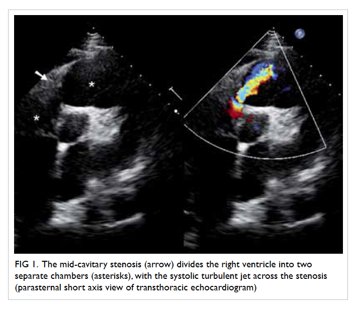
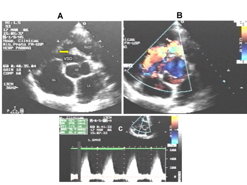
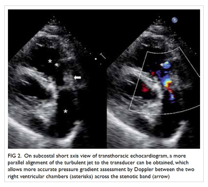
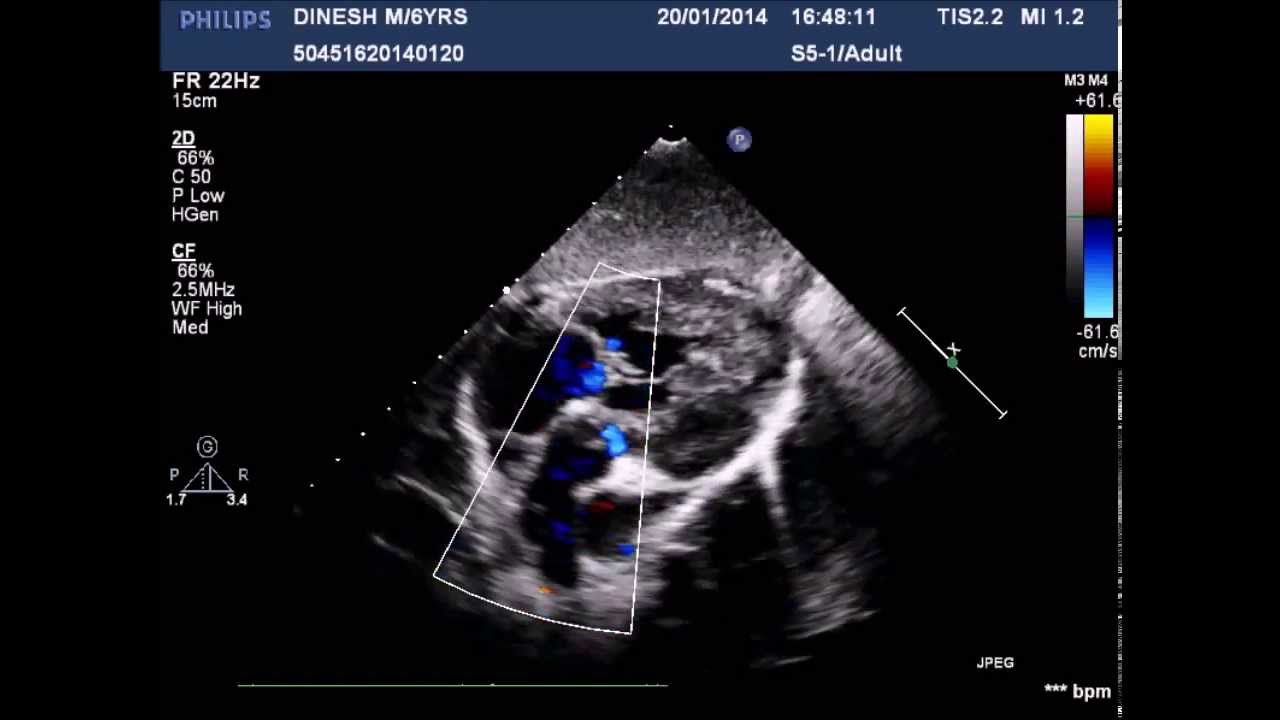
1. Appreciate abnormal right ventricular muscle bundle (RVMB) in apical four chamber and subcostal as well as parasternal views. Two-dimensional echo shows narrowing of mid RV cavity and color flow Doppler confirm the obstruction by displaying the turbulent jet. The subcostal view provides better alignment for measuring the pressure gradient by.

Double Outlet Right Ventricle (DORV)CausesSymptomsTreatment
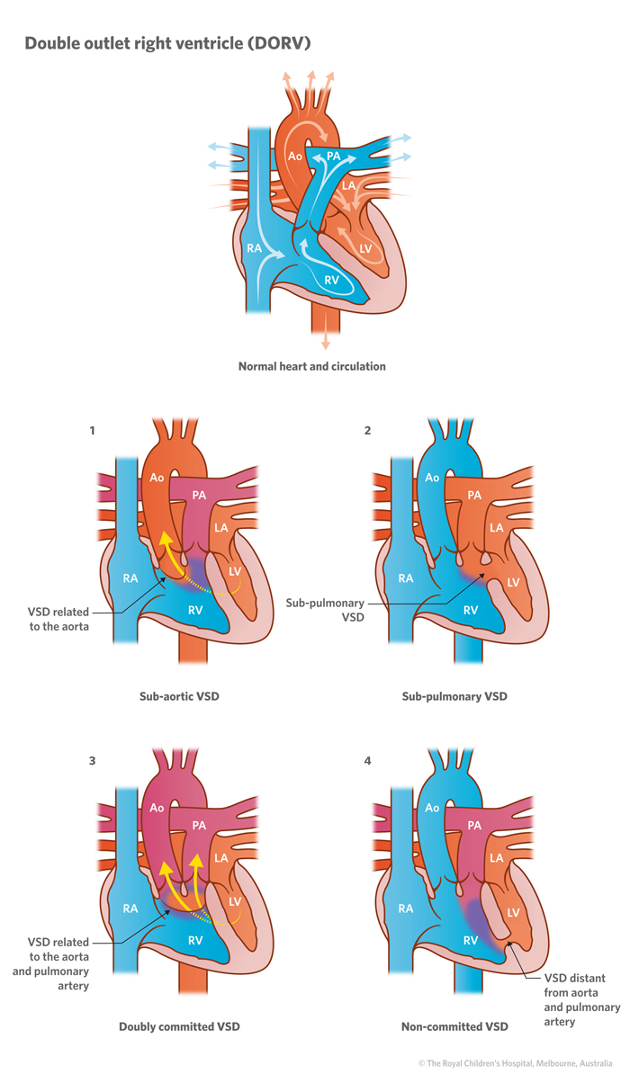
Double-outlet right ventricle is a heart condition present at birth. That means it's a congenital heart defect. In this condition, the body's main artery and the lung artery do not connect to the usual areas in the heart. The body's main artery is called the aorta. The lung artery is called the pulmonary artery.
An Adult With a DoubleChamber Right Ventricle Revista Española de
Double outlet right ventricle (DORV) is an abnormal heart condition in which two major arteries (instead of one) connect to your right ventricle or heart chamber. This is a congenital heart condition, which means you're born with it. Usually, each of your major blood vessels or "great" arteries connects to one of your heart's two ventricles.

9 best Double Chamber Right Ventricle (DCRV) images on Pinterest Baby
Double-chambered right ventricle (DCRV) occurs in approximately 1% of patients with congenital heart disease. The right ventricle (RV) is divided by anomalous muscle bundles into a higher-pressure proximal chamber and lower-pressure distal chamber. The physiology is defined by right ventricular pressure overload of the RV inflow chamber.

Pin by nonas arc on Double Outlet Right Ventricle Cardiac nursing
Fluid retention causing swelling in the lower limbs and sometimes the abdomen is a common and obvious symptom of right-sided heart failure. Still, there are several other symptoms that may develop.
Doublechambered right ventricle in an adult patient diagnosed by
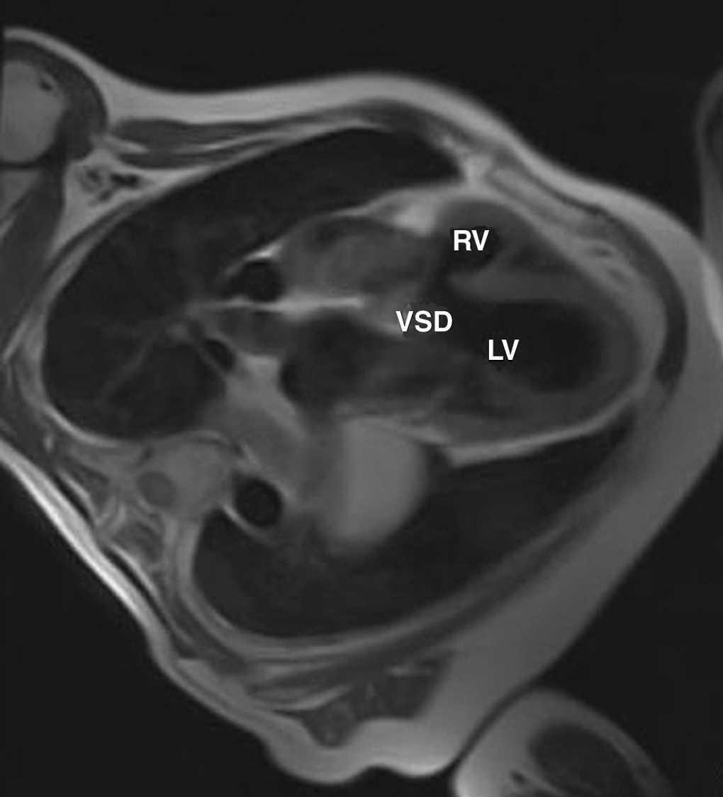
Double-chambered right ventricle is a rare congenital or acquired cardiac abnormality and may be associated with other malformations including membranous ventricular septal defect or double outlet right ventricle. 1 Patients may present with symptoms resembling ischemia or heart failure, including dyspnea and acute drops in blood pressure with s.
Doublechambered right ventricle a commonly overlooked diagnosis HKMJ
Double-chambered right ventricle is better understood as a form of septated right ventricle (RV) caused by the presence of abnormally located or hypertrophied muscular bands. The.
Double chambered right ventricle demonstrated by echocardiograghy YouTube
Double-chambered right ventricle (DCRV) was first described in 1858 by TB Peacock, but it is now understood to be a form of congenital heart disease wherein there is a mid-cavitary obstruction that divides the right ventricle into a high-pressure proximal portion and a low-pressure distal portion. D. Double-Chambered Right Ventricle Book

Figure 4 from Doublechambered right ventricle in a dog. Semantic Scholar
Double-chambered right ventricle (DCRV) is a cardiac disease of the right ventricular outflow tract obstruction characterized by anomalous muscle bundles (AMB) that divide the right ventricle into two chambers, a high-pressure inflow chamber and a low-pressure outflow chamber. The origin of AMB has been debated [ 1 - 3 ].

DoubleChambered Right Ventricle Circulation
Double outlet right ventricle surgery is a procedure that fixes a type of heart malformation called double outlet right ventricle (DORV). The normal heart has 4 chambers: 2 atria (upper chambers) and 2 ventricles (lower chambers). Blood flows from the right atrium into the right ventricle and from the left atrium into the left ventricle.
Cureus Adult DoubleChambered Right Ventricle Associated With
Double-chambered right ventricle (DCRV) is an uncommon congenital malformation in which anomalous muscle bundles dissect the RV into two chambers. It is commonly associated with other congenital anomalies, most frequently perimembranous ventricular septal defect (PM-VSD).

Figure 1 from Double Chambered Right Ventricle with Ventricular Septal
Tools Pill Identifier Formulary Recommended Like many other lesions associated with congenital heart disease (CHD), the terminology that surrounds double-chambered right ventricle (DCRV) has.